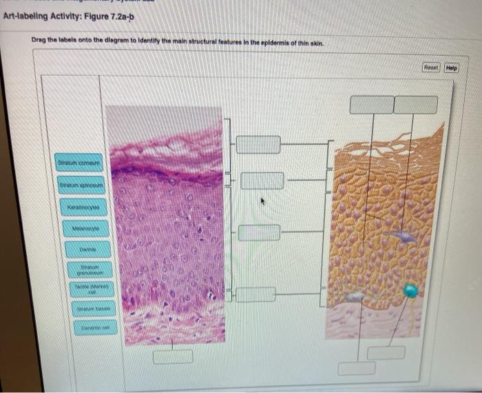
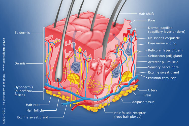
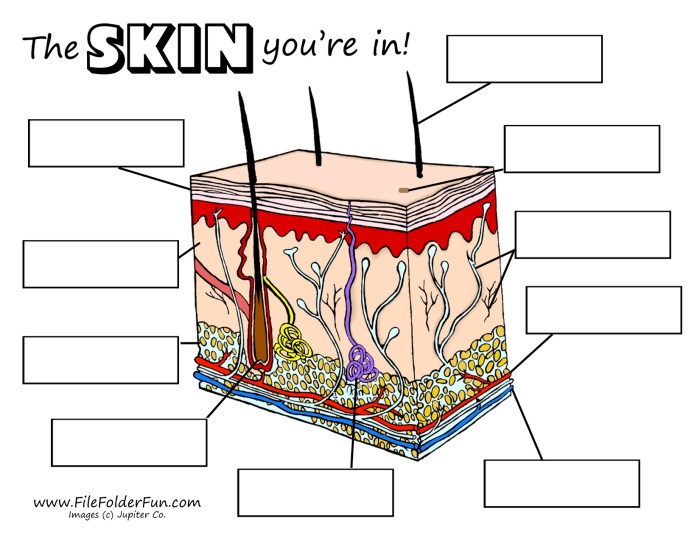
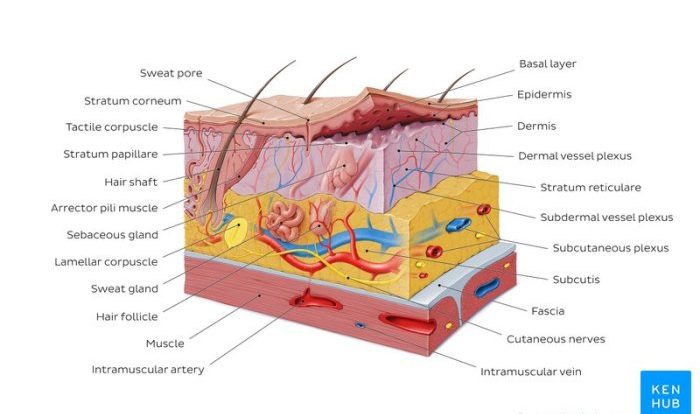
Art-labeling activity basic anatomy of the skin, an engaging and educational endeavor, invites you to explore the intricate layers of your skin, unveiling its remarkable structure and functions. Through a captivating blend of art and science, this activity illuminates the fundamental components of our largest organ, providing a deeper understanding of its essential role in maintaining our overall well-being.
Embark on a journey of discovery as we dissect the skin’s three distinct layers, unraveling the mysteries of the epidermis, dermis, and hypodermis. Delve into the diverse cell types that constitute the epidermis, each playing a vital role in protecting and nourishing our skin.
Uncover the secrets of the dermis, where collagen and elastin weave a resilient framework, and blood vessels sustain the skin’s vitality. Finally, explore the insulating and protective properties of the hypodermis, ensuring our bodies remain shielded from external elements.
Basic Anatomy of the Skin: Art-labeling Activity Basic Anatomy Of The Skin

The skin, the largest organ of the human body, serves as a protective barrier against the external environment. It consists of three distinct layers: the epidermis, dermis, and hypodermis, each with unique structures and functions.
Epidermis
The outermost layer of the skin, the epidermis, is composed of stratified squamous epithelium, a multi-layered tissue that provides protection against mechanical stress and UV radiation.
- Keratinocytes: The predominant cell type, producing the protein keratin, which provides structural strength and water resistance.
- Melanocytes: Responsible for producing melanin, the pigment that gives skin its color and protects against UV damage.
- Langerhans cells: Immune cells that detect and present antigens to the immune system.
- Merkel cells: Tactile receptors that detect light touch.
Dermis, Art-labeling activity basic anatomy of the skin
The middle layer of the skin, the dermis, provides structural support and elasticity.
- Collagen fibers: Provide tensile strength and prevent tearing.
- Elastin fibers: Provide elasticity and allow the skin to stretch and recoil.
- Blood vessels: Supply oxygen and nutrients to the skin.
- Hair follicles: Produce hair shafts.
- Sweat glands: Regulate body temperature through sweating.
- Sebaceous glands: Secrete sebum, which lubricates the skin and hair.
Hypodermis
The innermost layer of the skin, the hypodermis, consists of adipose tissue and connective tissue.
- Adipose tissue: Insulates the body and stores energy.
- Connective tissue: Provides structural support and anchors the skin to underlying structures.
Quick FAQs
What are the benefits of using art-based activities for teaching anatomy?
Art-based activities provide a unique and engaging way to learn anatomy by combining creativity with scientific concepts. They foster visual understanding, enhance spatial reasoning, and promote active recall, making the learning process more effective and memorable.
How can art-labeling activities be used to assess student understanding of the skin’s anatomy?
Art-labeling activities can serve as formative assessments, allowing educators to gauge students’ comprehension of skin anatomy. By observing students’ ability to accurately label and describe skin structures, educators can identify areas where further support or clarification is needed.
What are some examples of skin structures that can be included in an art-labeling activity?
Examples of skin structures that can be incorporated into an art-labeling activity include hair follicles, sweat glands, sebaceous glands, blood vessels, collagen fibers, and nerve endings. These structures represent the diverse components of the skin and provide a comprehensive overview of its anatomy.


